Joint dislocation
This article needs more reliable medical references for verification or relies too heavily on primary sources. (January 2022) |  |
| Joint dislocation | |
|---|---|
| Other names | Latin: luxatio |
 | |
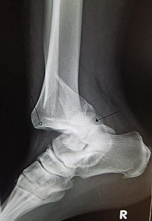
| A traumatic dislocation of the tibiotarsal joint of the ankle with distal fibular fracture. Open arrow marks the tibia and the closed arrow marks the talus. | |
| Specialty | Orthopedic surgery |
A joint dislocation, also called luxation, occurs when there is an abnormal separation in the joint, where two or more bones meet.[1] A partial dislocation is referred to as a subluxation. Dislocations are commonly caused by sudden trauma to the joint like during a car accident or fall. A joint dislocation can damage the surrounding ligaments, tendons, muscles, and nerves.[2] Dislocations can occur in any major joint (shoulder, knees, hips) or minor joint (toes, fingers). The most common joint dislocation is a shoulder dislocation.[1]
The treatment for joint dislocation is usually by closed reduction, that is, skilled manipulation to return the bones to their normal position. Only trained medical professionals should perform reductions since the manipulation can cause injury to the surrounding soft tissue, nerves, or vascular structures.[3]
Signs and Symptoms
[edit]The following symptoms are common with any type of dislocation.[1]
- Intense pain
- Joint instability
- Deformity of the joint area
- Reduced muscle strength
- Bruising or redness of the joint area
- Difficulty moving joint
- Stiffness
Complications
[edit]Joint dislocations can have associated injuries to surrounding tissues and structures, including muscle strains, ligament and tendon injuries, neurovascular injuries, and fractures.[4][5][6][7] Depending on the location of the dislocation, there are different complications to consider.
In the shoulder, vessel and nerve injuries are rare, but can cause many impairments and requires a longer recovery process.[4] Knee dislocations are rare, but can be complicated by injuries to arteries and nerves, leading to limb-threatening complications.[5] Degenerative changes following injury to the wrist are common, with many developing arthritis.[6] Persistent nerve pain years after the initial trauma is not uncommon.[6] Most finger dislocations occur in the middle of the finger (PIP) and are complicated by ligamentous injury (volar plate).[7] Since most dislocations involving the joint near the fingertip (DIP joint) are due to trauma, there is often an associated fracture or tissue injury.[7] Hip dislocations are at risk for osteonecrosis of the femoral head, femoral head fractures, the development of osteoarthritis, and sciatic nerve injury.[8][9] Given the strength of ligaments in the foot and ankle, ankle dislocation-fractures can occur.[10]
Causes
[edit]Joint dislocations are caused by trauma to the joint or when an individual falls on a specific joint.[11] Great and sudden force applied, by either a blow or fall, to the joint can cause the bones in the joint to be displaced or dislocated from their normal position.[12] With each dislocation, the ligaments keeping the bones fixed in the correct position can be damaged or loosened, making it easier for the joint to be dislocated in the future.[13]
Risk Factors
[edit]A variety of risk factors can predispose individuals to joint dislocation. They can vary depending on location of the joint. Genetic factors and underlying medical conditions can further increase risk. Genetic conditions, such as hypermobility syndrome and Ehlers-Danlos Syndrome put individuals at increased risk for dislocations.[14] Hypermobility syndrome is an inherited disorder that affects the ligaments around joints.[15] The loosened or stretched ligaments in the joint provide less stability and allow for the joint to dislocate more easily. Dislocation can also occur because of conditions such as Rheumatoid arthritis.[16] In Rheumatoid arthritis the production of synovial fluid decreases, gradually causing pain, swollen joints, and stiffness. A forceful push causes friction and can dislocate the joint. Notably, joint instability in the neck is a potential complication of rheumatoid arthritis.[16]
Participation in sports, being male, variations in the shape of the joint, being older, and joint hypermobility in males are risk factors associated with an increased risk of first time dislocation.[17]
Participation in sports, being a young male, history of a previous dislocation with an associated injury, and any history of previous dislocation are risk factors associated with recurrent dislocations.[17]
Diagnosis
[edit]Initial evaluation of a suspected joint dislocation begins with a thorough patient history, including mechanism of injury, and physical examination. Special attention should be focused on the neurovascular exam both before and after reduction, as injury to these structures may occur during the injury or during the reduction process.[3] Imaging studies are frequently obtained to assist with diagnosis and to determine the extent of injury.

Imaging Types
[edit]- Generally, pre- and post-reduction X-rays are taken. Initial X-ray can confirm the dislocation and evaluate for any fractures. Post-reduction x-rays confirm successful joint alignment and can identify any injuries that may have been caused during the reduction procedure.[18]
- If initial X-rays are normal but additional injury is suspected, there may be a benefit of obtaining stress/weight-bearing views to look for injury to ligamentous structures and/or need for surgical intervention. One example is with AC joint separations.[19]
- Ultrasound may be useful in an acute setting, and is a bedside test that can be performed in the Emergency Department. Ultrasound accuracy is dependent on user ability and experience. Ultrasound is nearly as effective as x-ray in detecting shoulder dislocations.[20][21] Ultrasound may also have utility in diagnosing AC joint dislocations.[22]
- In infants <6 months of age with suspected developmental dysplasia of the hip (congenital hip dislocation), ultrasound is the imaging study of choice. This is due to the lack of ossification at this age, which will not be apparent on x-rays. [23]
- X-rays are generally sufficient in confirming a joint dislocation. However, additional imaging can be used to better define and evaluate abnormalities that may be missed or unclear on plain X-rays. CT is not routinely used for simple dislocation, however it is useful in certain cases such as hip dislocation where an occult femoral neck fracture is suspected .[24] CT angiogram may be used if vascular injury is suspected.[24] In addition to improved visualization of bony abnormalities, MRI permits for a more detailed inspection of the joint-supporting structures in order to assess for ligamentous and other soft tissue injury.
Classification
[edit]Dislocations can either be full, referred to as luxation, or partial, referred to as subluxation. Simple dislocations are dislocations without an associated fracture, while complex dislocations have an associated fracture.[24] Depending on the type of joint involved (i.e. ball-and-socket, hinge), the dislocation can further be classified by anatomical position, such as an anterior hip dislocation.[24] Joint dislocations are named based on the distal component in relation to the proximal one.[25]
Prevention
[edit]Avoiding positions and activities that place the joint at risk for dislocation are effective strategies to prevent dislocation.[26] Similarly, wearing appropriate protective equipment during contact sports can be helpful. Strengthening exercises targeting muscles surrounding the joint are important to prevent dislocation.[26]
Treatment
[edit]Non-operative
[edit]Reduction/Repositioning
[edit]X-rays are taken to confirm a diagnosis and detect any fractures which may also have occurred at the time of dislocation. A dislocation is easily seen on an X-ray.[27] Once X-rays are taken, the joint is usually manipulated back into position. This can be a very painful process. This is typically done either in the emergency department under sedation or in an operating room under a general anaesthetic.[28] A dislocated joint should be reduced into its normal position only by a trained medical professional. Trying to reduce a joint without any training could worsen the injury.[29]
It is important to reduce the joint as soon as possible. Delaying reduction can compromise the blood supply to the joint. This is especially true in the case of a dislocated ankle, due to the anatomy of the blood supply to the foot.[30] On field reduction is crucial for joint dislocations. As they are extremely common in sports events, managing them correctly at the game at the time of injury, can reduce long term issues. They require prompt evaluation, diagnosis, reduction, and post-reduction management before the person can be evaluated at a medical facility.[31] After a dislocation, injured joints are usually held in place by a splint (for straight joints like fingers and toes) or a bandage (for complex joints like shoulders).
Immobilization
[edit]Immobilization is a method of treatment to place the injured joint in a sling or in another immobilizing device in order to keep the joint stable.[31] A 2012 Cochrane review, found no statistically significant difference in healing or long-term joint mobility between simple shoulder dislocations treated conservatively versus surgically.[32] Shorter immobilization periods are encouraged, with the goal of return to increased range-of-motion activities as soon as possible. [33][34] Shorter immobilization periods is linked to increased ranges of motion in some joints.[34]
Rehabilitation
[edit]Muscles, tendons and ligaments around the joint should be strengthened. This is usually done through a course of physical therapy, which will also help reduce the chances of repeated dislocations of the same joint[35] The most common treatment method for a dislocation of the Glenohumeral Joint (GH Joint/Shoulder Joint) is exercise based management.[36] For glenohumeral instability, the therapeutic program depends on specific characteristics of the instability pattern, severity, recurrence and direction with adaptations made based on the needs of the patient. In general, the therapeutic program should focus on restoration of strength, normalization of range of motion and optimization of flexibility and muscular performance. Throughout all stages of the rehabilitation program, it is important to take all related joints and structures into consideration.[37]
Operative
[edit]Surgery is often considered in extensive injuries or after failure of conservative management with strengthening exercises.[26] The need for surgery will depend on the location of the dislocation and the extent of the injury. Shoulder injuries can also be surgically stabilized, depending on the severity, using arthroscopic surgery.[27]
Prognosis
[edit]Prognosis varies depending on the location and extent of the dislocation. The prognosis of a shoulder dislocation is dependent on various factors including age, strength, connective tissue health and severity of the injury causing the dislocation.[24] There is a good prognosis in simple elbow dislocations in younger people. Older people report more pain and stiffness on average.[24] Wrist dislocations are often difficult to manage due to the difficulty in healing the small bones in the wrist.[24] Finger displacement towards the back of the hand is often irreducible due to associated injuries, while finger displacement towards the palm of the hand is more readily reducible.[24]
Epidemiology
[edit]Each joint in the body can be dislocated, however, there are common sites where most dislocations occur. The most common dislocated parts of the body are discussed as follows:
- Dislocated shoulder
- Anterior shoulder dislocation is the most common type of shoulder dislocation, accounting for at least 90% of shoulder dislocations. [4] [38] Anterior shoulder dislocations have a recurrence rate around 39%, with younger age at initial dislocation, male sex, and joint hyperlaxity being risk factors for increased recurrence.[39]
- The incidence rate of anterior shoulder dislocations is roughly 23.1 to 23.9 per 100,000 person-years.[40][41] Young males have a higher incidence rate, roughly four times that of the overall population.[40]
- Recurrent anterior shoulder dislocations have a higher rate of labrum tears (Bankart lesion) and humerus fractures/dents (Hill-Sachs lesion) compared to initial dislocations.[42]
- Shoulder dislocations account for 45% of all dislocation visits to the emergency room.[4]
- Elbow
- Wrist
- Finger
- Interphalangeal (IP) or metacarpophalangeal (MCP) joint dislocations[45]
- In the United States, men are most likely to sustain a finger dislocation with an incidence rate of 17.8 per 100,000 person-years.[46] Women have an incidence rate of 4.65 per 100,000 person-years.[46] The average age group that sustain a finger dislocation are between 15 and 19 years old.[46]
- The most common dislocations are in the proximal interphalangeal (PIP) joints.[7]
- Interphalangeal (IP) or metacarpophalangeal (MCP) joint dislocations[45]
- Hip
- Posterior and anterior hip dislocation
- Anterior dislocations are less common than posterior dislocations. 10% of all dislocations are anterior and this is broken down into superior and inferior types.[47] Superior dislocations account for 10% of all anterior dislocations, and inferior dislocations account for 90%.[47] 16-40 year old males are more likely to receive dislocations due to a car accident.[47]
- When an individual receives a hip dislocation, there is an incidence rate of 95% that they will receive an injury to another part of their body as well.[47]
- 46–84% of hip dislocations occur secondary to traffic accidents, the remaining percentage is due based on falls, industrial accidents or sporting injury.[39]
- Posterior and anterior hip dislocation
- Knee
- The majority of knee dislocations (64.5%) are caused by trauma to the knee, with more than half caused by car and motorcycle accidents.[48]
- The incidence rate of initial patellar dislocations is roughly 32.8 per 100,000 person years.[41]
- Nearly 41% of knee dislocations have an associated fracture, with the majority of these fractures in one of the legs.[48]
- Nerve injury occurs in about 15.3% of knee dislocations, while major artery injury occurs in 7.8% of knee dislocations.[48]
- More than half (53.5%) of knee dislocations have an associated torn meniscus.[48]
- Tendon rupture occurs up to 13.1% of the time.[48]
- Foot and Ankle
- A lisfranc injury is a dislocation or fracture-dislocation injury at the tarsometatarsal joints.
- A subtalar dislocation, or talocalcaneonavicular dislocation, is a simultaneous dislocation of the talar joints at the talocalcaneal and talonavicular levels.[49][50]
- Subtalar dislocations without associated fractures represent about 1% of all traumatic injuries of the foot and 1-2% of all dislocations, and they are caused by high energy trauma. Early closed reduction is recommended, otherwise open reduction without further delay.[51]
- A total talar dislocation is rare with high rates of complications.[52][53]
- Ankle Sprains primarily occur as a result of tearing the ATFL (anterior talofibular ligament) in the Talocrural Joint. The ATFL tears most easily when the foot is in plantarflexion and inversion.[54]
- An ankle dislocation without fracture is rare, due to the strength of ligaments surrounding the ankle.[55]
Gallery
[edit]-
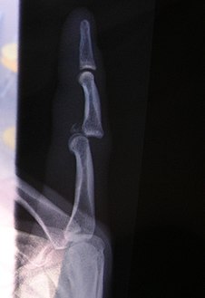
Dislocation of the left index finger
-
Radiograph of right fifth phalanx bone dislocation
-
Radiograph of left index finger dislocation
-
Depiction of reduction of a dislocated spine, ca. 1300
-
Dislocation of the carpo-metacarpal joint.
-
Radiograph of right fifth phalanx dislocation resulting from bicycle accident
-
Right fifth phalanx dislocation resulting from bicycle accident
-
Shoulder dislocation before (left) and after (right) being reduced
See also
[edit]- Buddy wrapping
- Major trauma
- Physical therapy
- Projectional radiography
- Listhesis, olisthesis, or olisthy
References
[edit]- ^ a b c "Dislocations". Lucile Packard Children’s Hospital at Stanford. Archived from the original on 28 May 2013. Retrieved 3 March 2013.
- ^ Smith RL, Brunolli J (February 1989). "Shoulder kinesthesia after anterior glenohumeral joint dislocation". Physical Therapy. 69 (2): 106–112. doi:10.1093/ptj/69.2.106. PMID 2913578.
- ^ a b Skelley NW, McCormick JJ, Smith MV (May 2014). "In-game Management of Common Joint Dislocations". Sports Health. 6 (3): 246–255. doi:10.1177/1941738113499721. PMC 4000468. PMID 24790695.
- ^ a b c d Khiami F, Gérometta A, Loriaut P (February 2015). "Management of recent first-time anterior shoulder dislocations". Orthopaedics & Traumatology, Surgery & Research. 101 (1 Suppl): S51 – S57. doi:10.1016/j.otsr.2014.06.027. PMID 25596982.
- ^ a b Gómez-Bermúdez SJ, Vanegas-Isaza D, Herrera-Almanza L, Roldán-Tabares MD, Coronado-Magalhaes G, Fernández-Lopera JF, et al. (2021). "[Vascular injury associated with knee dislocation]". Acta Ortopedica Mexicana. 35 (2): 226–235. doi:10.35366/101872. PMID 34731929.
- ^ a b c d e Heineman N, Do DH, Golden A (September 2023). "Carpal dislocations". The Journal of Hand Surgery, European Volume. 48 (2_suppl): 11S – 17S. doi:10.1177/17531934231183260. PMID 37704022.
- ^ a b c d Borchers JR, Best TM (April 2012). "Common finger fractures and dislocations". American Family Physician. 85 (8): 805–810. PMID 22534390.
- ^ Masiewicz S, Mabrouk A, Johnson DE (2025). "Posterior Hip Dislocation". StatPearls. Treasure Island (FL): StatPearls Publishing. PMID 29083669. Retrieved 23 January 2025.
- ^ Graber M, Marino DV, Johnson DE (2025). "Anterior Hip Dislocation". StatPearls. Treasure Island (FL): StatPearls Publishing. PMID 29939591.
- ^ Lawson KA, Ayala AE, Morin ML, Latt LD, Wild JR (July 2023). "Republication of "Ankle Fracture-Dislocations: A Review"". Foot & Ankle Orthopaedics. 8 (3): 24730114231195058. doi:10.1177/24730114231195058. PMC 10423454. PMID 37582190.
- ^ "Finger Dislocation Joint Reduction". Mayo Clinic.
- ^ "Dislocation". U.S. National Library of Medicine.
- ^ "Dislocation – Joint dislocation". Pubmed Health.
- ^ "Ehlers-Danlos syndromes". Genetic and Rare Diseases Information Center (GARD) – an NCATS Program. 24 September 2017. Retrieved 16 January 2025.
- ^ Ruemper A, Watkins K (December 2012). "Correlations Between General Joint Hypermobility and Joint Hypermobility Syndrome and Injury in Contemporary Dance Students". Journal of Dance Medicine & Science : Official Publication of the International Association for Dance Medicine & Science. 16 (4): 161–6. PMID 26731093.
- ^ a b Subagio EA, Wicaksono P, Faris M, Bajamal AH, Abdillah DS (5 October 2023). Dalal V (ed.). "Diagnosis and Prevalence (1975-2010) of Sudden Death due to Atlantoaxial Subluxation in Cervical Rheumatoid Arthritis: A Literature Review". TheScientificWorldJournal. 2023: 6675489. doi:10.1155/2023/6675489. PMC 10569890. PMID 37841539.
- ^ a b Wright A, Ness B, Spontelli-Gisselman A, Gosselin D, Cleland J, Wassinger C (1 May 2024). "Risk Factors Associated with First Time and Recurrent Shoulder Instability: A Systematic Review". International Journal of Sports Physical Therapy. 19 (5): 522–534. doi:10.26603/001c.116278. PMC 11065770. PMID 38707855.
- ^ Chong M, Karataglis D, Learmonth D (September 2006). "Survey of the management of acute traumatic first-time anterior shoulder dislocation among trauma clinicians in the UK". Annals of the Royal College of Surgeons of England. 88 (5): 454–458. doi:10.1308/003588406X117115. PMC 1964698. PMID 17002849.
- ^ Gaillard F. "Acromioclavicular injury". Radiology Reference Article. Radiopaedia.org. Retrieved 21 February 2018.
- ^ Abbasi S, Molaie H, Hafezimoghadam P, Zare MA, Abbasi M, Rezai M, et al. (August 2013). "Diagnostic accuracy of ultrasonographic examination in the management of shoulder dislocation in the emergency department". Annals of Emergency Medicine. 62 (2): 170–175. doi:10.1016/j.annemergmed.2013.01.022. PMID 23489654.
- ^ Gottlieb M, Patel D, Marks A, Peksa GD (August 2022). "Ultrasound for the diagnosis of shoulder dislocation and reduction: A systematic review and meta-analysis". Academic Emergency Medicine. 29 (8): 999–1007. doi:10.1111/acem.14454. PMID 35094451.
- ^ Heers G, Hedtmann A (June 2005). "Correlation of ultrasonographic findings to Tossy's and Rockwood's classification of acromioclavicular joint injuries". Ultrasound in Medicine & Biology. 31 (6): 725–732. doi:10.1016/j.ultrasmedbio.2005.03.002. PMID 15936487.
- ^ Gaillard F (2 May 2008). "Developmental dysplasia of the hip". Radiology Reference Article. Radiopaedia.org. Retrieved 21 February 2018.
- ^ a b c d e f g h i Tornetta P, ed. (2020). Rockwood and Green's fractures in adults (9th ed.). Philadelphia: Wolters Kluwer. ISBN 978-1-4963-8651-9.
- ^ "Introduction to Trauma X-ray - Dislocation injury". www.radiologymasterclass.co.uk. Retrieved 15 February 2018.
- ^ a b c McMahon PJ (2021). Current Diagnosis & Treatment in Orthopedics (6th ed.). McGraw-Hill Education.
- ^ a b Dias JJ, Steingold RF, Richardson RA, Tesfayohannes B, Gregg PJ (November 1987). "The conservative treatment of acromioclavicular dislocation. Review after five years". The Journal of Bone and Joint Surgery. British Volume. 69 (5): 719–22. doi:10.1302/0301-620X.69B5.3680330. PMID 3680330.
- ^ Holdsworth F (December 1970). "Fractures, dislocations, and fracture-dislocations of the spine". The Journal of Bone and Joint Surgery. American Volume. 52 (8): 1534–51. PMID 5483077.
- ^ Bankart AB (July 1938). "The pathology and treatment of recurrent dislocation of the shoulder-joint". Journal of British Surgery. 26 (101): 23–29. doi:10.1002/bjs.18002610104.
- ^ Ganz R, Gill TJ, Gautier E, Ganz K, Krügel N, Berlemann U (November 2001). "Surgical dislocation of the adult hip a technique with full access to the femoral head and acetabulum without the risk of avascular necrosis". The Journal of Bone and Joint Surgery. British Volume. 83 (8): 1119–1124. doi:10.1302/0301-620x.83b8.11964. PMID 11764423.
- ^ a b Skelley NW, McCormick JJ, Smith MV (May 2014). "In-game Management of Common Joint Dislocations". Sports Health. 6 (3): 246–255. doi:10.1177/1941738113499721. PMC 4000468. PMID 24790695.
- ^ Taylor F, Sims M, Theis JC, Herbison GP, et al. (Cochrane Bone, Joint and Muscle Trauma Group) (April 2012). "Interventions for treating acute elbow dislocations in adults". The Cochrane Database of Systematic Reviews. 2012 (4): CD007908. doi:10.1002/14651858.CD007908.pub2. PMC 6465046. PMID 22513954.
- ^ Barco R, Gonzalez-Escobar S, Acerboni-Flores F, Vaquero-Picado A (November 2023). "Acute elbow dislocation: a critical appraisal of the literature". JSES International. 7 (6): 2560–2564. doi:10.1016/j.jseint.2023.03.019. PMC 10638560. PMID 37969505.
- ^ a b Breulmann FL, Lappen S, Ehmann Y, Bischofreiter M, Lacheta L, Siebenlist S (February 2024). "Treatment strategies for simple elbow dislocation - a systematic review". BMC Musculoskeletal Disorders. 25 (1): 148. doi:10.1186/s12891-024-07260-0. PMC 10874000. PMID 38365699.
- ^ Itoi E, Hatakeyama Y, Kido T, Sato T, Minagawa H, Wakabayashi I, et al. (2003). "A new method of immobilization after traumatic anterior dislocation of the shoulder: a preliminary study". Journal of Shoulder and Elbow Surgery. 12 (5): 413–5. doi:10.1016/s1058-2746(03)00171-x. PMID 14564258.
- ^ Warby SA, Pizzari T, Ford JJ, Hahne AJ, Watson L (January 2014). "The effect of exercise-based management for multidirectional instability of the glenohumeral joint: a systematic review". Journal of Shoulder and Elbow Surgery. 23 (1): 128–142. doi:10.1016/j.jse.2013.08.006. PMID 24331125.
- ^ Cools AM, Borms D, Castelein B, Vanderstukken F, Johansson FR (February 2016). "Evidence-based rehabilitation of athletes with glenohumeral instability". Knee Surgery, Sports Traumatology, Arthroscopy. 24 (2): 382–389. doi:10.1007/s00167-015-3940-x. PMID 26704789. S2CID 21227767.
- ^ Breed M, Fitch RW (2021). The Atlas of Emergency Medicine (5th ed.). McGraw-Hill.
- ^ a b Olds M, Ellis R, Donaldson K, Parmar P, Kersten P (July 2015). "Risk factors which predispose first-time traumatic anterior shoulder dislocations to recurrent instability in adults: a systematic review and meta-analysis". British Journal of Sports Medicine. 49 (14): 913–922. doi:10.1136/bjsports-2014-094342. PMC 4687692. PMID 25900943.
- ^ a b Olds M, Ellis R, Donaldson K, Parmar P, Kersten P (July 2015). "Risk factors which predispose first-time traumatic anterior shoulder dislocations to recurrent instability in adults: a systematic review and meta-analysis". British Journal of Sports Medicine. 49 (14): 913–922. doi:10.1136/bjsports-2014-094342. PMC 4687692. PMID 25900943.
- ^ a b c Ponkilainen V, Kuitunen I, Liukkonen R, Vaajala M, Reito A, Uimonen M (November 2022). "The incidence of musculoskeletal injuries: a systematic review and meta-analysis". Bone & Joint Research. 11 (11): 814–825. doi:10.1302/2046-3758.1111.BJR-2022-0181.R1. PMC 9680199. PMID 36374291.
- ^ Rutgers C, Verweij LP, Priester-Vink S, van Deurzen DF, Maas M, van den Bekerom MP (June 2022). "Recurrence in traumatic anterior shoulder dislocations increases the prevalence of Hill-Sachs and Bankart lesions: a systematic review and meta-analysis". Knee Surgery, Sports Traumatology, Arthroscopy. 30 (6): 2130–2140. doi:10.1007/s00167-021-06847-7. PMC 9165262. PMID 34988633.
- ^ Hackl M, Beyer F, Wegmann K, Leschinger T, Burkhart KJ, Müller LP (May 2015). "The treatment of simple elbow dislocation in adults". Deutsches Arzteblatt International. 112 (18): 311–319. doi:10.3238/arztebl.2015.0311. PMC 4455254. PMID 26037467.
- ^ Halstead ME, Bernhardt DT, Garry JP (16 November 2022). Talavera F, Goitz HT, Young CC (eds.). "Elbow Dislocation". Medscape.
- ^ Polansky R, Kwon NS (27 June 2023). Windle ML, Lovato LM, Schraga ED (eds.). "Reduction of Finger Dislocation". Medscape.
- ^ a b c Golan E, Kang KK, Culbertson M, Choueka J (June 2016). "The Epidemiology of Finger Dislocations Presenting for Emergency Care Within the United States". Hand. 11 (2): 192–196. doi:10.1177/1558944715627232. PMC 4920528. PMID 27390562.
- ^ a b c d Clegg TE, Roberts CS, Greene JW, Prather BA (April 2010). "Hip dislocations--epidemiology, treatment, and outcomes". Injury. 41 (4): 329–334. doi:10.1016/j.injury.2009.08.007. PMID 19796765.
- ^ a b c d e Randall ZD, Strok MJ, Mazzola JW, Agrawal R, Yaeger LH, Berkes MB (November 2024). "The known and unknown reality of knee dislocations: A systematic review". Injury. 55 (11): 111904. doi:10.1016/j.injury.2024.111904. PMID 39357194.
- ^ Ruhlmann F, Poujardieu C, Vernois J, Gayet LE (2017). "Isolated Acute Traumatic Subtalar Dislocations: Review of 13 Cases at a Mean Follow-Up of 6 Years and Literature Review". The Journal of Foot and Ankle Surgery (Review). 56 (1): 201–207. doi:10.1053/j.jfas.2016.01.044. PMID 26947001. S2CID 31290747.
- ^ García-Regal J, Centeno-Ruano AJ (2013). "[Talocalcaneonavicular dislocation without associated fractures]". Acta Ortopedica Mexicana (Review) (in Spanish). 27 (3): 201–204. PMID 24707608.
- ^ Prada-Cañizares A, Auñón-Martín I, Vilá Y Rico J, Pretell-Mazzini J (May 2016). "Subtalar dislocation: management and prognosis for an uncommon orthopaedic condition". International Orthopaedics (Review). 40 (5): 999–1007. doi:10.1007/s00264-015-2910-8. PMID 26208589. S2CID 6090499.
- ^ Foy MA, Fagg PS (5 December 2011). Medicolegal Reporting in Orthopaedic Trauma E-Book. Elsevier Health Sciences. pp. 320–. ISBN 978-0-7020-4886-9.
- ^ For a graphic representation of displacements that may lead to a total talar dislocation see: Bucholz RW (29 March 2012). Rockwood and Green's Fractures in Adults: Two Volumes Plus Integrated Content Website (Rockwood, Green, and Wilkins' Fractures). Lippincott Williams & Wilkins. p. 2061. ISBN 978-1-4511-6144-1.
- ^ Ringleb SI, Dhakal A, Anderson CD, Bawab S, Paranjape R (October 2011). "Effects of lateral ligament sectioning on the stability of the ankle and subtalar joint". Journal of Orthopaedic Research. 29 (10): 1459–1464. doi:10.1002/jor.21407. PMID 21445995.
- ^ Wight L, Owen D, Goldbloom D, Knupp M (October 2017). "Pure Ankle Dislocation: A systematic review of the literature and estimation of incidence". Injury (Review). 48 (10): 2027–2034. doi:10.1016/j.injury.2017.08.011. PMID 28826653.
- ^ Earwaker J (1992). "Posttraumatic calcification of the annular ligament of the radius". Skeletal Radiology. 21 (3): 149–154. doi:10.1007/BF00242127. PMID 1604339. S2CID 43615869.
External links
[edit]


![X-ray of ventral dislocation of the radial head. There is calcification of annular ligament, which can be seen as early as 2 weeks after injury.[56]](http://upload.wikimedia.org/wikipedia/commons/6/66/X-ray_of_ventral_dislocation_of_the_radial_head_with_calcification_of_annular_ligament.jpg)
